For Material Science
For Biological Science
Sampling Tools and Techniques for Material Science
Material preparation Laboratory
Our unit has a well equipped lab that contains all the chemicals and equipments required for Material sample preparation.
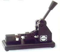 The preferred method for preparing disks from ductile materials such as metals. This disc punch is specifically designed to avoid compression and distortion of the sample. It cuts a disc size of 3mm (2.3mm) diameter and material thickness ranging from 10 µm to above 100 µm.
 Prethins and polishes specimens for shorter ion milling times.
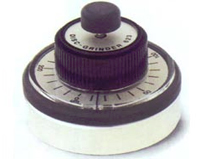
 This grinder will reduce, with minimal damage, the central region of a typical 100µm thick and 3mm diameter specimen blank to a few micrometers in times ranging from 20 minutes for silicon to 100 minutes for sapphire. Subsequent chemical or particle beam thinning is then completed rapidly to produce large electron transparent areas.
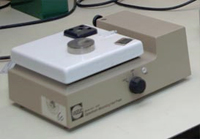 It is most important that specimens are firmly attached during ultrasonic cutting. This is best done by using a low melting point wax polymer to form a strong, thin, hard adhesive band. The specimen mounting hotplate is thermostatically controlled at the precise mounting temperature of 130°C. The hotplate also contains recesses to hold the specimen mounts in place when bounding the discs.
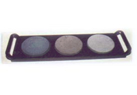 This kit consists of a heavy metal base, 3 ultra-flat glass lapping plates and 60 adhesive lapping discs. The metal base holds the glass plates in place as the disc grinder is moved with a figure-eight action over the lapping discs. 40µm, 15µm and 5µm grit are used in sequence to produce highly polished specimens.
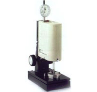 The disc cutter is gentle enough to cut discs from wafers as thin as 50µm and also powerful enough to cut 5 mm long cylinders from bulk materials in only minutes. The cutting tool is driven by a crystal piezo (lead zirconate / itanate) at a frequency of 28 KHZ. The cutting medium consists of a water based slurry of silicon or boron carbide. A constant tool load is automatically maintained during cutting to give the maximum erosion rate attainable without risking thermal or microstructure damage to the TEM specimen, the depth of cut is displayed continually on a dial indicator (10µm resolution) which is mechanically coupled to the specimen platform.
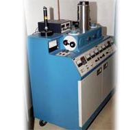 Ion beam milling is now the preferred method for preparing TEM specimens of ceramics, semi conductors and metals. Ion beam milling is done by dual ion guns. Ion beams aimed at glancing angles of incidence to the specimen i.e. ~ 12°. Low angle thinning has the advantage that it produces larger thin areas of specimen. While minimizing beam heating and radiation damage effects, the machine is equipped with a low powered optical microscope that can be situated directly above the specimen. The process of ion beam thinning will be automatically terminated as the specimen perforates by Laser Auto terminators. Also variable speed specimen rotation is available.
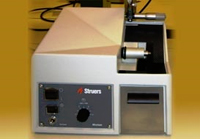
Small, automatic precision cut-off machine for sectioning all materialographic and ceramic specimens.
Minitom is very easy to operate, with fixation of the specimen and the setting of specimen size, cutting speed, and cutting pressure taking only a few minutes. The workpiece is fastened in a holder mounted on a movable arm.
Minitom is a small, low-speed, precision cut-off machine for sectioning all types of materials. Maximum specimen size: 30 mm dia. The cutting speed is continuously variable and the motor is designed to ensure that the selected speed remains constant at any load.
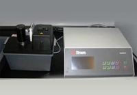
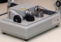
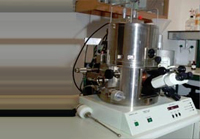
Apparatus for automatic, electrolytical thinning of specimens for examination in a transmission electron microscope. With the built-in scanning function and additional enhancement functions parameters for new materials can easily be established.
Specimens with a diameter of 2.3 and 3 mm dia are polished from both sides simultaneously to achieve a thin foil with a centre hole as small as possible. The thinning is controlled by an infrared light source, stopping the process as soon as the first light can pass through the established hole.
Sampling Tools and Techniques for Biological Science
Biological preparation Laboratory
Our unit has a well equipped lab that contains all the chemicals and equipments required for Biological sample preparation
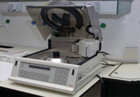
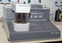
This tissue processor is used for automatic fixation, dehydration and monomer infiltration of biological materials. It is safe to use, reliable and economic. Processing capacity specifications as follows:
1. olds up to 56 specimens.
2. Holds up to 7 baskets for 4 0r 8 small specimens or 3 baskets for 3 large specimens.
3. Program steps can be individually controlled from 1 minute to 99 hours 59 minutes.
4. Up to 10 programs may be stored with selection of time, temperature and agitation for each of the 20 steps.
5. Programmable delay starts of program.
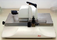
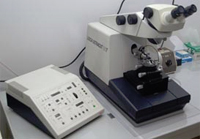
Reichert Glass Knife maker to produce high Quality triangular 45° glass Knives for Ultramicrotomy with long Cutting edge from special 6, 4, 8 and 10mm, thick glass.

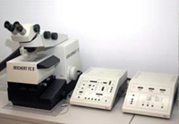
This new and most modern Ultramicrotome is simple in operation. In the routine, specimen ultrathin and semi thin sectioning for transmission electron microscopy is done. It can be found with cryosectianing system.
These are the basic features:
Basic instrument with 180° rotating microscope carrier, multi illumination system for 7 different illuminations, stepping motor for 200µm advance, motor for 10 mm knife N-S movement and drive for 25 mm E-W movement. Reflomat II waterpump, knife block 360° rotatable, drive graduated at ± 30°, clearance angle drive -2° to +15°, knife holder for 6-12 mm knives, segment are 360° rotatable, self-locking precision drives for ± 22° eucentric rotating +47° +3° setting and the 360° rotation of the specimen holder. Trimming block, trimming adapter R, control unit with speed control 0.05' to 100 mm/sec, 3 click stops return speed 10/20/30 mm/sec, dual automatic advance with dual feed setting, ultra 0-95 nm, semi 0.01 - 2.5µm, cutting window control 0.1 - 14 mm, warning 20µm before the end, advance Reset control, Joystick for knife movement, built-in section counter and feed totalizer. Available with universal specimen holder UT, flat specimen holder FT, stereo microscope BL with 200 m objective 0.67 - 4.0 x, wide field eyepieces WF 15 X..
This new and most modern Ultramicrotome is simple in operation. In the routine, specimen ultrathin and semi thin sectioning for transmission electron microscopy is done. It can be found with cryosectianing system.
These are the basic features :
Basic instrument with 180° rotating microscope carrier, multi illumination system for 7 different illuminations, stepping motor for 200µm advance, motor for 10 mm knife N-S movement and drive for 25 mm E-W movement. Reflomat II waterpump, knife block 360° rotatable, drive graduated at ± 30°, clearance angle drive -2° to +15°, knife holder for 6-12 mm knives, segment are 360° rotatable, self-locking precision drives for ± 22° eucentric rotating +47° +3° setting and the 360° rotation of the specimen holder. Trimming block, trimming adapter R, control unit with speed control 0.05' to 100 mm/sec, 3 click stops return speed 10/20/30 mm/sec, dual automatic advance with dual feed setting, ultra 0-95 nm, semi 0.01 - 2.5µm, cutting window control 0.1 - 14 mm, warning 20µm before the end, advance Reset control, Joystick for knife movement, built-in section counter and feed totalizer.Available with universal specimen holder UT, flat specimen holder FT, stereo microscope BL with 200 m objective 0.67 - 4.0 x, wide field eyepieces WF 15 X..
E.M. Grid stainer for safe and automatic double staining of ultra thin sections with sealed and stabilized air free packed stains. Ultra thin sections are automatically stained to yield high-contrast, precipitate free specimens. By staining several grids simultaneously, the ultrastainer speeds up laboratory routines and eliminates the many tedious tasks associated with orthodox traditional staining methods. Because the ultrastainer uses prepacked stains, there is no need to prepare toxic uranyl and lead solutions. Ultrastain I and II are vacuum sealed to ensure precipitate-free and safe staining. The microprocessor of ultrastainer is programmed for a basic staining sequence suitable for most applications. For special staining, however, it is possible to set your own parameters.
This system is used for the dry pumping of the cryo holder of the cryo transfer system and double tilt cryo holder for JEM-1200 EX II TEM.
1. Biological specimens intended for investigation in the Scanning electron microscope must be dried before being introduced to the microscope. If specimens are dried in air, tangential forces caused by the molecular linkage force of the liquid at the liquid-air interface affect the minuscule structures protruding from the liquid. The resulting surface tension deforms the structure of the specimen, and can even destroy it entirely.
2. Critical point drying is a method in which the liquid goes directly into the gas phase (no freezing first), but deforming forces are avoided because the drying process takes place above the critical point of the liquid where the phase boundary between liquid and gas no longer exists.
3. Critical point dryer - CPD-030 - Features:
4. Compact table top Model.
5. Good specimen visibility from above and laterally through generously dimensioned observation port.
6. Vertical loading with quick-closing lid.
7. Automatic pre cooling.
8. Built-in heating, electronically regulated and continuously adjustable up to 50°C.
9. Certified Pressure Chamber (200 bar) with safety bursting membrane (150 bar) integrated magnetic stirrer.
10. Simple and safe operation for all conventional dehydrating agents.
11. Separate inlets and outlets for coolant and dehydrating agents.
Etching device, e.g. for eliminating detrimental absorbed oxygen. Simple switchover between sputtering and etching. Adjustable specimen height. Pre selection of the sputtering time with electronics. Built in solenoid valve for interrupting the flow of argon after the sputtering unit has terminated Automatic venting valve.
Etching device, e.g. for eliminating detrimental absorbed oxygen. Simple switchover between sputtering and etching. Adjustable specimen height. Pre selection of the sputtering time with electronics. Built in solenoid valve for interrupting the flow of argon after the sputtering unit has terminated Automatic venting valve.
State of the art electronics with digital displays for vacuum stage temperature, sputtering time, either sputter voltage or current and for current drawn during carbon evaporation.
All operational controls in the form of a touch-pad keyboard.
Safety of operator assured due to newly designed protective device incorporating two vacuum sensors and a safety interlock. Quickly replaced foil targets.
1 Freeze Fraction and Freeze Etching.
The financial grant for this lab is given by KFAS. It is equipped with JFD-9010 freeze etching and other supporting equipment for sample preparation.
The JFD-9010 freeze etching equipment is used to prepare specimens for electron microscopy by various treatments of fresh biological specimens, frozen by rapid freezing. The treatment includes freeze-fracturing and freeze-etching in high vacuum for maintaining cells and tissues of such specimens in an almost native state, plus electron beam evaporation for making replica films of ultra - micro particles.
Also, use of electron beam evaporator and specimen rotation/tilt mechanism allows low-angle rotational evaporation to be performed with ease. This is extremely useful for molecular biologists, who employ electron microscope for high-resolution observation of protein molecules.
2 Cryoultramicrotomy.
1. Sectioning down to-185°C.
2. New contact-free through-the-wall specimen holder system for chatter free cryosection.
3. Open top design with no disturbing ice-condensation inside or outside during or after work.
4. Attaches to the Ultracut S and at low temperature within a few minutes.
5. Memory for 3x3 settings of knife specimen and gas temperature. Continuous turbulence-free no-pressure LN2filling system with a very low LN2 consumption.
6. New shell mounted sectioning chamber with armrest for comfortable manipulation of sections.
7. Accurate control of specimen, knife and the most important chamber gas temperature.
8. Backlight illumination for accurate glass and safe diamond knife approach.
9. Alignment of knife with self-locking drives from outside the chamber.
3 Cryotransferring system.
 This system, with Cryo-holder, ultra thin sections grids, can be loaded directly from Cryoultramicrotome to cryo holder in the microtomy room. Then, it can be transferred at low temperature to the Transmission electron microscope.